Tendon Injuries: Flexor & Extensor Tendons
Complete guide to finger tendon injuries including mallet finger, jersey finger, boutonniere deformity, and surgical repair techniques.
Understanding Tendon Anatomy
Flexor Tendons: Two flexor tendons to each finger - Flexor Digitorum Superficialis (FDS) and Flexor Digitorum Profundus (FDP). FDP flexes the DIP joint; FDS flexes the PIP joint. They run through a pulley system (A1-A5) that keeps them close to bone. The "no man's land" (Zone II) between the A1 pulley and FDS insertion is particularly challenging for repair.
Extensor Tendons: More superficial and flat. The extensor mechanism is complex, with the central slip inserting at the middle phalanx and lateral bands joining to insert at the distal phalanx. Injuries at different levels create distinct deformities.
Zones: Flexor tendons have 5 zones (I-V) and extensor tendons have 8 zones (I-VIII). Zone location affects treatment approach and prognosis.
Common Tendon Injuries
Extensor tendon avulsion at DIP joint. Fingertip droops, cannot extend actively.
Treatment: Splint DIP in extension 6-8 weeks (continuous). Surgery only for large bony fragments or failed splinting.
FDP avulsion from distal phalanx. Cannot flex DIP. Ring finger most common.
Treatment: Surgical repair required. Ideally within 7-10 days. Outcomes depend on tendon retraction level.
Central slip rupture. PIP flexion, DIP hyperextension. Often from PIP dislocation or laceration.
Treatment: Early - splint PIP extended. Late/fixed - may need surgical reconstruction.
Opposite of boutonniere - PIP hyperextension, DIP flexion. Multiple causes including volar plate injury, RA.
Treatment: Depends on cause and flexibility. Splinting, soft tissue procedures, or fusion.
Signs & Symptoms
• Loss of movement: Cannot actively flex or extend specific joint
• Deformity: Characteristic postures (mallet, boutonniere, swan neck)
• Pain: At injury site, especially with attempted movement
• Swelling: Over affected area
• Weakness: Reduced grip or pinch strength
• Visible laceration: In open injuries
Clinical Testing: FDP function - hold finger at PIP, ask patient to flex DIP. FDS function - hold other fingers extended, ask patient to flex PIP. Extensor function - extend each joint against resistance.
Diagnosis
Clinical Examination: The cornerstone of diagnosis. Test each tendon individually:
- FDP: Stabilize finger at PIP, have patient flex DIP
- FDS: Hold all other fingers extended, have patient flex isolated finger at PIP
- Extensors: Extend each joint against resistance
- Cascade: With wrist extended, fingers should progressively flex from index to small
X-rays: Check for bony avulsions (mallet, jersey finger). Rule out associated fractures or dislocations.
Ultrasound/MRI: Can confirm diagnosis and show tendon retraction level. Useful when clinical exam is difficult.
Treatment Principles
Appropriate for specific injuries:
- Mallet finger (tendon only): DIP extension splint for 6-8 weeks continuous, then night splinting
- Acute boutonniere: PIP extension splint for 6 weeks with DIP exercises
- Partial tendon lacerations (<50%): May heal with splinting
Key: Compliance is critical. Removing the splint "just for a minute" resets the clock.
Indications:
- Complete tendon lacerations
- Jersey finger (FDP avulsion)
- Open injuries with tendon involvement
- Large bony mallet with subluxation
- Failed non-surgical treatment
Surgical Techniques:
Primary Repair: Direct suturing of tendon ends. Best outcomes when done early. Various suture techniques (modified Kessler, cruciate, etc.)
Tendon Grafting: For gaps or late presentation. Palmaris longus or plantaris used as graft.
Two-Stage Reconstruction: For scarred bed. First stage: silicone rod creates new sheath. Second stage: tendon graft.
Tenolysis: Release of adhesions around healed but stiff tendon. Done after fractures heal.
Rehabilitation Protocols
Kleinert Protocol: Early passive flexion with rubber band traction, active extension. Dynamic splint.
Duran Protocol: Controlled passive motion. No active motion for 4 weeks.
Early Active Motion: Place-and-hold exercises. Requires strong repair.
Full motion by 8-12 weeks. Strengthening after 12 weeks.
Zones I-II: DIP splinting 6-8 weeks.
Zones III-IV: PIP extension splinting, early DIP motion.
Zones V-VIII: Dynamic extension splinting, early motion protocols.
Extensors more forgiving than flexors for adhesions.
Key Point: Hand therapy is essential after tendon repair. The balance between protecting the repair and preventing adhesions is critical. Protocol compliance directly affects outcomes.
Evidence & References
- • Strickland JW. Development of flexor tendon surgery: twenty-five years of progress. J Hand Surg Am. 2000
- • Doyle JR. Extensor tendons - acute injuries. Green's Operative Hand Surgery. 2017
- • Tang JB. Clinical outcomes associated with flexor tendon repair. Hand Clin. 2005
- • Handoll HH, Vaghela MV. Interventions for treating mallet finger injuries. Cochrane Database Syst Rev. 2004
Last Updated: January 2025 | Content reviewed by Dr. Deepthi Nandan Reddy, FRCS (Ortho)
Frequently Asked Questions
YOUR TREATING SURGEON
Dr. Deepthi Nandan Reddy
Senior Consultant Orthopaedic & Upper-Limb Surgeon | Apollo Hospitals
Learn From Dr. Reddy
Watch expert explanations of conditions and treatment options
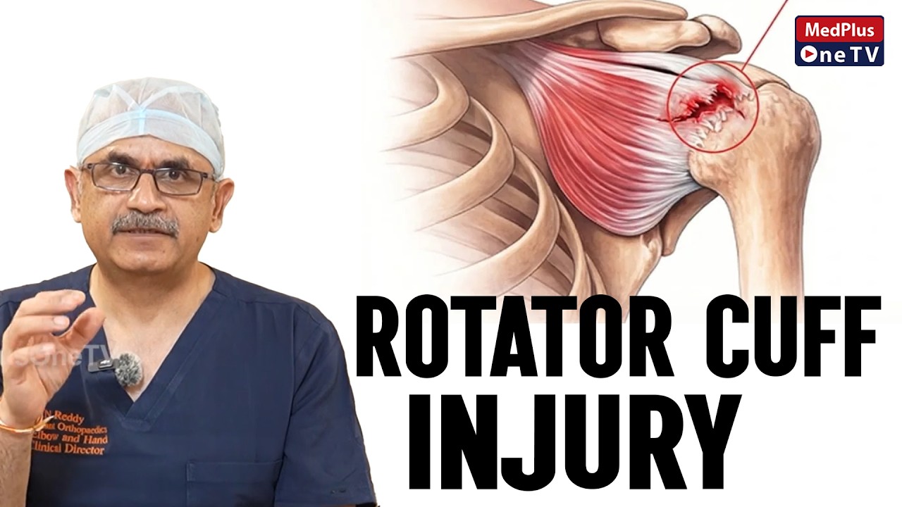
Understanding Rotator Cuff Injuries

Tennis Elbow Pain - Causes & Treatment
Shoulder Instability Explained
Why Choose Dr. Deepthi Nandan Reddy?
International fellowship training combined with decades of experience delivering world-class upper limb care
UK CCT
Certificate of Completion of Training - One of few Indian surgeons with NHS Consultant experience
FRCS (UK)
Fellow of Royal College of Surgeons, Edinburgh & UK
Mayo Clinic Fellow
British Shoulder & Elbow Society - Mayo Clinic Travelling Fellowship
SESI President
President, Shoulder & Elbow Society of India
30+ Years Experience
15,000+ patients treated, 5,000+ surgeries performed
Published Researcher
Co-author of Operative Elbow Surgery, international conference faculty
Dr. Reddy's Specialty Clinics
Comprehensive Upper Limb Care
Continue Learning