Peripheral Nerve Injuries of the Hand
Complete guide to median, ulnar, and radial nerve injuries. Understanding diagnosis, surgical repair, nerve transfers, and rehabilitation.
Understanding Nerve Injuries
Peripheral nerve injuries can result in loss of sensation, muscle paralysis, and significant functional impairment. The three main nerves serving the hand - median, ulnar, and radial - each have specific sensory and motor functions. Understanding these patterns is crucial for diagnosis and treatment planning.
Nerve Anatomy: Peripheral nerves consist of bundles (fascicles) of nerve fibers surrounded by protective layers. Motor and sensory fibers may be mixed or grouped. This architecture influences repair techniques and outcomes.
Regeneration: Unlike the central nervous system, peripheral nerves can regenerate. After injury, the nerve distal to the injury degenerates (Wallerian degeneration), then slowly regrows from the injury site at approximately 1mm/day.
Sunderland Classification
Neurapraxia
Conduction block. No structural damage. Full recovery in days-weeks.
Axonotmesis
Axon damage, tubes intact. Full recovery expected. 1mm/day.
Endoneurial damage
Variable recovery. Some misdirection of regrowing fibers.
Perineurial damage
Only epineurium intact. Surgery usually needed.
Neurotmesis
Complete transection. Surgical repair required.
Major Nerve Injuries
Sensory loss: Thumb, index, middle, radial half of ring finger (palmar)
Motor loss: Thenar muscles (thumb opposition, abduction) - "ape hand" deformity
High injury: Also affects forearm flexors - loss of thumb IP and index DIP flexion
Common causes: Lacerations at wrist, supracondylar fractures, carpal tunnel syndrome
Sensory loss: Small finger and ulnar half of ring finger
Motor loss: Intrinsic muscles - "claw hand" deformity (worse in low injuries)
Weakness: Grip, pinch strength, finger abduction/adduction
Froment's sign: Thumb IP flexion during pinch (FPL substitution for adductor)
Common causes: Lacerations, cubital tunnel, Guyon's canal compression
Sensory loss: Dorsal hand (first web space) - often minimal functional impact
Motor loss: Wrist and finger extension - "wrist drop"
High injury: Also affects triceps and brachioradialis
Common causes: Humeral shaft fractures, "Saturday night palsy", posterior interosseous nerve syndrome
Diagnosis
Clinical Examination:
- Sensory testing: Light touch, two-point discrimination (normal <6mm)
- Motor testing: Test specific muscles innervated by each nerve
- Tinel's sign: Percussion over nerve produces tingling - tracks regeneration
- Provocative tests: Phalen's (median), Froment's (ulnar)
Electrodiagnostic Studies (EMG/NCS):
- Perform 3-4 weeks post-injury (time for Wallerian degeneration)
- Helps localize injury level
- Differentiates complete from incomplete injuries
- Monitors recovery - serial studies at 3-month intervals
Imaging: MRI neurography for complex cases. Ultrasound can show nerve continuity and neuromas.
Treatment Options
Appropriate for closed injuries with potential for spontaneous recovery:
- Observation with serial clinical and EMG evaluation
- Splinting to prevent joint contracture
- Hand therapy to maintain ROM
- Sensory re-education once recovery begins
If no recovery by 3-4 months, surgical exploration indicated.
Primary Nerve Repair
Direct suturing of nerve ends. Best when done early (within 72 hours for sharp injuries). Tension-free repair essential. Epineurial or group fascicular repair techniques.
Nerve Grafting
When gap prevents tension-free repair. Sural nerve (leg) most common donor. Bridging graft allows regeneration through conduit. Expect some loss compared to direct repair.
Nerve Transfer
Sacrificing less important nerve to power more important function. Shortens regeneration distance for proximal injuries. Examples: AIN to ulnar motor, median to radial transfer.
Tendon Transfer (Salvage)
When nerve repair not possible or has failed. Working muscles redirected to replace paralyzed function. Can restore excellent function. Examples: FCU to finger extensors for radial palsy.
Recovery & Rehabilitation
Sensory Re-education: Systematic training to help the brain interpret altered sensory signals. Begins when protective sensation returns. Involves texture recognition, object identification, and localization exercises.
Evidence & References
- • Sunderland S. A classification of peripheral nerve injuries producing loss of function. Brain. 1951
- • Mackinnon SE, Dellon AL. Surgery of the Peripheral Nerve. 1988
- • Ray WZ, Mackinnon SE. Management of nerve gaps: autografts, allografts, nerve transfers, and end-to-side neurorrhaphy. Exp Neurol. 2010
- • Novak CB, et al. Evaluation of nerve injury and nerve repair. Hand Clin. 2013
Last Updated: January 2025 | Content reviewed by Dr. Deepthi Nandan Reddy, FRCS (Ortho)
Frequently Asked Questions
YOUR TREATING SURGEON
Dr. Deepthi Nandan Reddy
Senior Consultant Orthopaedic & Upper-Limb Surgeon | Apollo Hospitals
Learn From Dr. Reddy
Watch expert explanations of conditions and treatment options
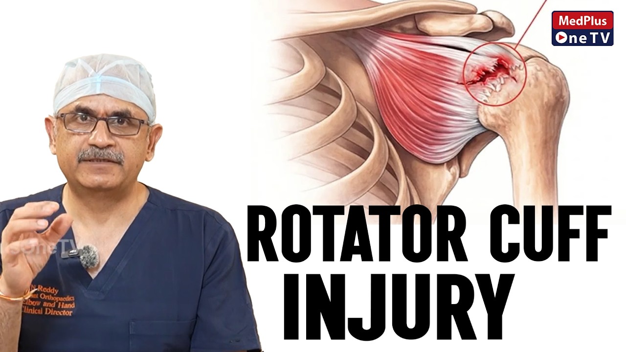
Understanding Rotator Cuff Injuries
Tennis Elbow Pain - Causes & Treatment
Shoulder Instability Explained
Why Choose Dr. Deepthi Nandan Reddy?
International fellowship training combined with decades of experience delivering world-class upper limb care
UK CCT
Certificate of Completion of Training - One of few Indian surgeons with NHS Consultant experience
FRCS (UK)
Fellow of Royal College of Surgeons, Edinburgh & UK
Mayo Clinic Fellow
British Shoulder & Elbow Society - Mayo Clinic Travelling Fellowship
SESI President
President, Shoulder & Elbow Society of India
30+ Years Experience
15,000+ patients treated, 5,000+ surgeries performed
Published Researcher
Co-author of Operative Elbow Surgery, international conference faculty
Dr. Reddy's Specialty Clinics
Comprehensive Upper Limb Care
Continue Learning